
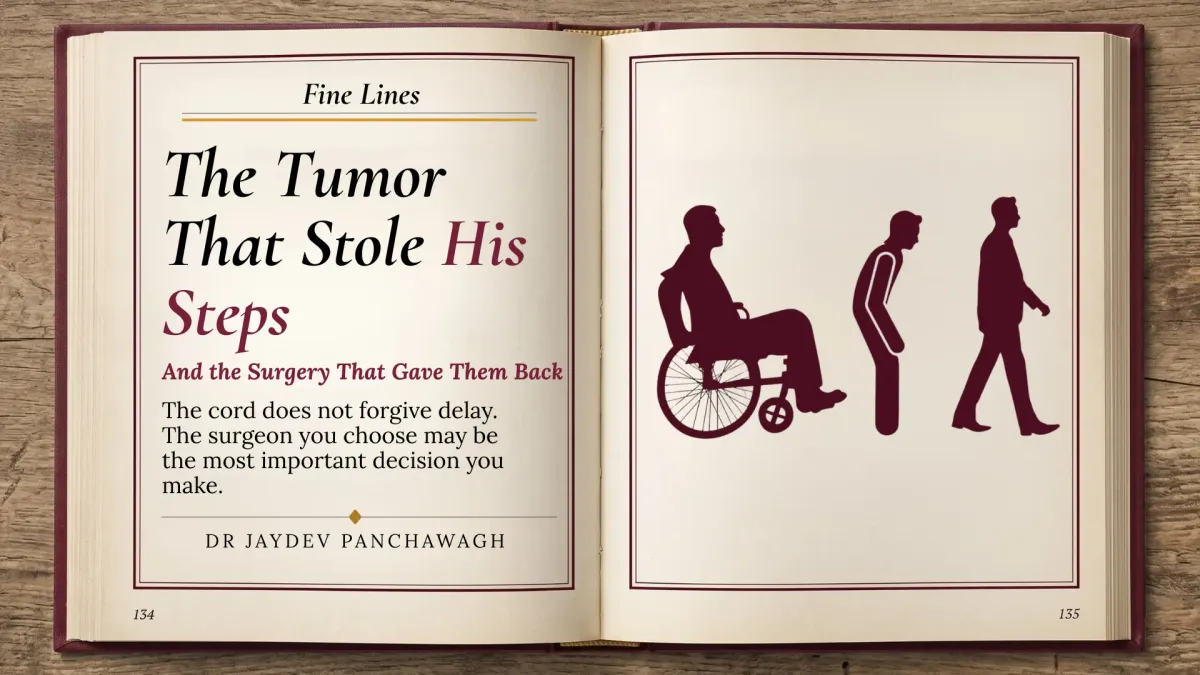
The cord does not forgive delay. The surgeon you choose may be one of the most important decisions you make.
— Dr. Jaydev Panchawagh · Neurosurgeon, Pune
The Anatomy
What was growing inside — how a spinal cord tumour can announce itself, and why its early signs may be missed
Some patients enter a clinic and the diagnosis is already written in how they move.
Mr. Pradeep was forty-four years old when he first noticed something was wrong. A civil contractor, accustomed to long days on construction sites and longer evenings on his feet, he dismissed the ache between his shoulder blades as the price of a physical life. His legs, he told himself, were tired. The stumble on a flat surface — that was nothing. The heaviness in his thighs — that was age, or overwork, or both.
But the symptoms progressed. Over the following weeks, heaviness became weakness and weakness became unreliability. One morning, several months after the symptoms began, Mr. Pradeep could not rise from his bed without his wife’s help.
He arrived in a wheelchair, pushed by a son who looked more frightened than his father.
The spinal cord is among the most precisely organised structures in the human body. It carries signals between the brain and the body — instructions for movement, sensations such as touch and pain, and pathways involved in bladder and bowel function. It runs through a protective canal of bone and membranes, with limited space around it.
When a tumour grows within the spinal canal, it can gradually compress the cord. Early symptoms may be easy to mistake for something ordinary: a dull back ache, fatigue in the legs, tingling or subtle changes in balance. These complaints may initially resemble a disc problem, overwork or age-related discomfort.
This is one challenge with spinal cord compression: symptoms can develop gradually and be mistaken for less serious conditions. By the time weakness affects standing or walking, the cord may have been under pressure for some time. Timely assessment matters, although the extent of recovery varies from patient to patient.
The Diagnosis
The MRI and what it told us — why the tumour’s position matters
Mr. Pradeep’s MRI showed a well-defined mass inside his spinal canal at the thoracic level — the mid-back — sitting outside the spinal cord itself but pressing against it. The cord was visibly displaced to one side.
This was an intradural extramedullary spinal tumour: it lay within the dural sac, the membrane enclosing the cord and its surrounding fluid, but outside the cord’s own substance. This distinction matters when planning surgery and discussing prognosis. A tumour arising within the cord can present different surgical challenges from one that presses against it from outside.
Common tumours in this category include schwannomas, which arise from cells associated with peripheral nerve coverings, and meningiomas, which develop from the meninges. Many are benign, although their behaviour and treatment depend on the individual diagnosis, location and imaging features.
The tumour in Mr. Pradeep’s thoracic canal had severely affected his ability to walk. The MRI suggested that the cord was compressed rather than invaded by the mass. That distinction was important in planning the operation, while the degree and duration of compression remained relevant to the recovery discussion.
The Decision
The son’s homework — how the family considered experience, facilities and the surgical team
Mr. Pradeep’s son — a young engineer in his late twenties, methodical by temperament — spent two weeks after the MRI making phone calls.
He first contacted a nearby large hospital. The team could operate, he was told, but the surgeon had limited experience with intradural spinal cord tumours specifically. He called a second centre, two hours away. They were willing to treat his father, but did not have dedicated intraoperative neurophysiological monitoring — the real-time signals that can help the surgical team assess the function of neural pathways during an operation.
He then spoke to a physiotherapist who had observed outcomes across multiple centres. The advice was to ask about the surgeon’s experience with this specific type of tumour and whether the necessary operating microscope, monitoring and postoperative support were available. For complex spinal cord surgery, the relevant experience and the capabilities of the whole team matter.
The son made a longer journey. He arrived with his father in the wheelchair, a carefully organised folder of imaging and a list of questions written in the margins of a notebook.
I have met this kind of family before. They ask not only how soon an operation can be arranged, but how often the team performs this kind of surgery, what monitoring is used and what recovery may realistically involve. They understand that spinal cord surgery requires detailed familiarity with the anatomy and a team equipped for the procedure.
The operating microscope helps the surgeon distinguish the tumour from the compressed cord and surrounding structures. Learning to identify and follow the appropriate tissue plane takes specialised training and experience. The choice of centre should therefore consider the surgeon’s relevant experience, available technology, anaesthesia, monitoring and rehabilitation support — not convenience alone.
The Operation
Inside the operating theatre — microsurgical removal with neurological monitoring
We operated under general anaesthesia. The surgical approach opened the spinal canal from behind, giving access to the intradural space where the tumour was located.
Under the operating microscope, the anatomy that had appeared compressed and distorted on the MRI became clearer, layer by careful layer. The dura was opened, followed by the arachnoid membrane beneath it. The spinal cord was pale and displaced, with the tumour resting against it — well-defined and distinct from the cord tissue around it.
Neurophysiological monitoring was used throughout the procedure to help assess the function of relevant neural pathways. The tumour was carefully dissected away from the cord, progressing millimetre by millimetre while preserving surrounding structures.
There was no moment of drama. Good spinal cord surgery rarely has drama. It has patience, precision and familiarity with the anatomy.
The tumour was removed in its entirety. With the mass no longer compressing it, the cord was freed from the pressure that had affected it for months.
The Recovery
Standing up — the first steps after months of weakness
Over the years, I have come to treasure a particular moment in recovery after spinal cord tumour surgery. It is the moment when I ask the physiotherapist to help the patient stand.
There is often hesitation. Patients who have not walked in months, who have arrived in wheelchairs and who fear that their weakness may be permanent do not immediately trust their legs. They need encouragement, support and time.
Mr. Pradeep stood.
His legs were weak. The spinal cord does not reverse months of compression in a matter of days. Recovery after decompression can continue over weeks and months, and it depends on factors such as the cause and duration of compression, the severity of neurological changes before surgery and the patient’s rehabilitation.
He began walking with a walker and later progressed to a stick. Four months after arriving at the clinic in a wheelchair, he walked in on his own, with a steadier gait and a handshake that told me how much the progress meant to him.
His bladder function, which had been severely impaired before surgery, had returned. His bowel control had improved, and he was back at work. His recovery was deeply encouraging, though every patient’s outcome and timeline are different.
What This Means
A diagnosis is not a sentence — the factors that shape recovery after spinal cord tumour surgery
Surgery for selected benign intradural extramedullary tumours, including schwannomas and meningiomas, can be rewarding when the tumour can be safely separated from the cord and treated at an appropriate stage. Outcomes vary with the tumour type, its location and its relationship to the spinal cord and nerve roots.
Three factors are especially important when discussing what surgery may achieve. The type and position of the tumour influence the technical options. The spinal level involved brings its own anatomical considerations. And the degree of neurological impairment at the time of treatment helps shape how much function may recover.
These factors are not all within a patient’s control. But recognising progressive symptoms, arranging appropriate investigation and seeking a team with relevant experience can influence the care pathway.
The difficulty in spinal cord tumour cases is that early symptoms may be mistaken for tiredness, ordinary back pain or a familiar disc problem. New or progressive weakness in the legs, persistent numbness, changes in balance, or bladder and bowel symptoms deserve prompt medical assessment. A clinician may recommend MRI when the history and examination suggest possible spinal cord compression.
When an MRI reveals a spinal cord tumour, ask about the exact diagnosis, the proposed approach, the surgeon’s experience with similar tumours, the monitoring available and the rehabilitation plan.
Mr. Pradeep walked into the clinic four months after he had been wheeled in. His progress reflected the value of relieving the compression and giving rehabilitation time to work. The lesson is not that every patient will have the same result, but that progressive neurological symptoms should not be ignored.
A Note on Fear and Surgery
Understanding the risks of spinal cord tumour surgery without allowing fear to replace informed decisions
Patients facing spinal cord tumour surgery often arrive carrying a specific and understandable fear: that the operation will add to the damage the tumour has begun. The spinal cord is delicate, and the possibility of a new neurological deficit is a risk that must be discussed honestly.
The risk depends on the tumour’s type, size, position and relationship to the cord and nerve roots, as well as the patient’s existing neurological condition and the surgical team’s experience. An operating microscope and intraoperative neurophysiological monitoring can support careful surgery, but they cannot eliminate risk or guarantee a particular outcome.
Leaving a compressive tumour untreated may also carry risks, including further neurological deterioration. The appropriate balance between surgery, observation or another treatment depends on the individual diagnosis and should be discussed with the treating specialist.
Fear is not a reason to delay seeking advice. It is a reason to ask clear questions, understand the alternatives and make an informed decision with an experienced team.
Names and identifying details have been changed to protect patient privacy. Shared with the patient’s consent.
Continue Reading
When the Spine Speaks
Recognising symptoms that may signal a problem involving the spine and nervous system.
Read More →
The Face That Feared the Wind
A patient’s experience of facial pain, diagnosis and treatment for trigeminal neuralgia.
Read More →
The Pain Nobody Believed
Why persistent symptoms deserve careful evaluation and a search for their cause.
Read More →


