Introduction
Did you know that a jelly-like, fluid-filled cyst can silently develop inside your brain? This rare condition is called a Third Ventricular Colloid Cyst. If not detected in time, it can pose the serious risk of sudden death.
But here’s the good news: early diagnosis and timely surgery can lead to a completely normal, healthy life.
Dr. Malti Panchawagh, anesthesiologist at Brain and Spine Surgery Center led by India’s renowned Neurosurgeon Dr. Jaydev Panchwagh , explains this condition in simple language to help raise awareness—especially among non-medical audiences.
Understanding the Brain’s Ventricular System
To understand this cyst better, we must first understand the ventricular system of the brain. The brain has four chambers, or ventricles, filled with a vital fluid called Cerebrospinal Fluid (CSF).
CSF performs three main functions:
The ventricles are:
Lateral Ventricles (2): The largest ventricles, where CSF is produced
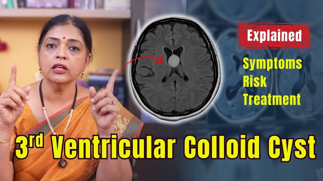
Third Ventricle: Receives CSF from the lateral ventricles
Fourth Ventricle: CSF flows from here to the spinal cord and brain surface
This fluid is continuously produced and circulates throughout the brain and spine before being absorbed back into the bloodstream.
What is a Colloid Cyst?
A colloid cyst is a slow-growing, non-cancerous structure filled with sticky fluid. Its wall gradually produces more fluid over time, causing the cyst to enlarge.
Key facts:
Accounts for only 2% of primary brain tumors
99% are located in the third ventricle
Most common in people aged 30–70, though can occur in children
Why It’s Dangerous: Symptoms and Sudden Death Risk
In early stages, small cysts may cause no symptoms. However, as they grow, they can block the flow of CSF, especially between the lateral and third ventricles, leading to a condition called hydrocephalus (fluid buildup in the brain).
Symptoms may include:
Persistent headaches
Nausea or vomiting
Blurred vision
Ringing in the ears (tinnitus)
Dizziness or imbalance
Memory loss
Confusion
Difficulty walking or unsteadiness
Brief unconsciousness
In severe cases: coma or even sudden death
Because of this risk, early detection and treatment are critical.
Diagnosis: MRI and CT Scans
Doctors use CT scans or MRI to determine:
The size and location of the cyst
Whether CSF flow is obstructed
The level of pressure on brain structures
MRI can also help visualize the CSF pathway and assess the risk more clearly.
Treatment Options: Two Types of Surgery
1. Endoscopic Removal
Minimally invasive procedure
A small cut and a tiny hole are made in the skull
A thin tube-like instrument (endoscope) is inserted to remove the cyst
Faster recovery, less pain, and minimal scarring
2. Conventional Craniotomy
Traditional open surgery
A larger incision is made
Surgeons access the cyst with microscopic precision tools
Used in complex cases or when endoscopy isn’t feasible
Both surgeries at Brain Surgery Center use AI-based navigation systems and neuro-precision tools for safe and accurate cyst removal.
Final Takeaways
Third Ventricular Colloid Cysts are non-cancerous but can be life-threatening if untreated.
They are rare, but dangerous due to their location in the brain.
Early diagnosis and timely surgery can restore a patient’s normal life.
Treatment options include endoscopic surgery or traditional craniotomy, based on individual needs.
Early diagnosis and advanced surgical options can help treat third ventricular colloid cysts safely.I
If you found this information helpful, please share it—it may help save a life.
Stay informed, stay healthy, and take care of your loved ones.


Leave a Reply